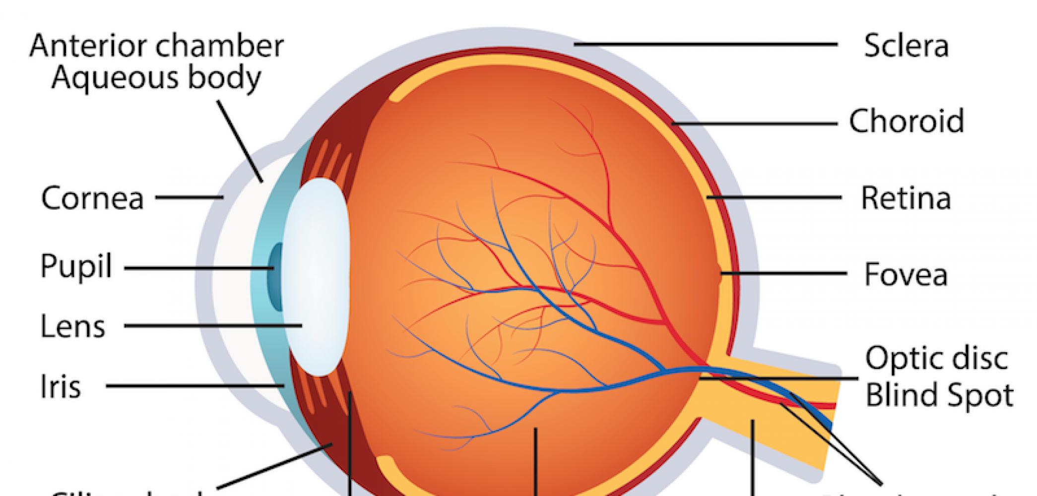
Basic Eye Anatomy And Physiology

Human Eye Anatomy La Pine Eyecare Clinic The surface of the eye and the inner surface of the eyelids are covered with a clear membrane called the conjunctiva. the layers of the tear film keep the front of the eye lubricated. tears lubricate the eye and are made up of three layers. these three layers together are called the tear film. the mucous layer is made by the conjunctiva. The eye is kept moist by secretions of the lacrimal glands (tear glands). these almond shaped glands under the upper lids extend inward from the outer corner of each eye. each gland has two portions. one portion is in a shallow depression in the part of the eye socket formed by the frontal bone.

Basic Anatomy And Physiology Of The Human Visual System Eye Anatomy Medical Anatomical The following are parts of the human eyes and their functions: 1. conjunctiva. the conjunctiva is the membrane covering the sclera (white portion of your eye). the conjunctiva also covers the interior of your eyelids. the conjunctiva helps lubricate the eyes by generating mucus and tears. it also aids in immunological monitoring and prevents. Your eyes are a key sensory organ, feeding information to your brain about the outside world. your eyes do the “physical” part of seeing. the signals they send allow your brain to “build” the picture that you see. eye related symptoms are also key clues to issues affecting your whole body, so experts recommend making eye health a priority. Learn about eye anatomy and learn how your eyes work with ophthalmologist approved facts. please note: this website includes an accessibility system. press control f11 to adjust the website to the visually impaired who are using a screen reader; press control f10 to open an accessibility menu. Dr. mike's briefly explains the layers of the eyeball:outer: sclera and corneamiddle: choroid, ciliary body, irisinner: retinaand also discusses the lens and.

Comments are closed.