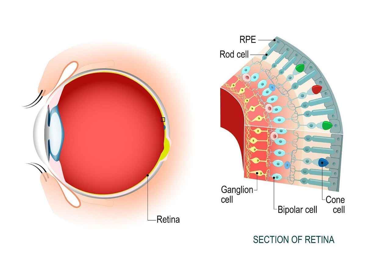
Human Eye Structure Image Formation And Difference Between Rods And Cones Simplebiology

Rods Cones In The Human Eye Retina is the sensitive layer as it has sensitive cells which help in the formation of image and differentiation of colours in the picture. retina has two types of cells: rods. cones. these cells help in the formation of the picture on the retina. cones separate colours while the rods are responsive to white light. The human eye has two main types of photoreceptors, rods and cones, which get their names from their shapes. rods. these photoreceptors are tall and have a cylindrical (tubelike) shape. they’re extremely sensitive to even tiny amounts of light. about 95% of the photoreceptors in your eyes (about 100 125 million) are rods.

Human Eye Structure Image Formation And Difference Between Rods And Cones Simplebiology The retina is the light sensitive part at the back of the eye. there are two photoreceptor types: rods and cones. signals from these photoreceptors are sent to the brain for processing via the optic nerve. the optic nerve is a bundle of nerve fibers that connects each eye’s retina to the brain. 1. there are more rod photoreceptors than cone. It supports the shape of the eye and provides enough distance so that the lens can focus. retina: the retina is the coating on the inside of the back of the eye. it contains two types of cells. rods detect light and help form images in dim light. cones detect colors. there are three types of cones. On that account, fovea centralis contains the highest number of cones in the retina, and has the highest visual acuity of the eye. similarities between rods and cones. both rods and cones are the photoreceptor cells in the vertebrae retina. both rods and cones contain visual pigments. both rods and cones are types of secondary exteroreceptor cells. The rods are photoreceptors that contain the visual pigment – rhodopsin and are sensitive to blue green light with a peak sensitivity around 500 nanometers (nm) wavelength of light (fig. 14a). rods are highly sensitive photoreceptors and are used for vision under dark dim conditions at night.

Human Eye Structure Image Formation And Difference Between Rods And Cones Simplebiology On that account, fovea centralis contains the highest number of cones in the retina, and has the highest visual acuity of the eye. similarities between rods and cones. both rods and cones are the photoreceptor cells in the vertebrae retina. both rods and cones contain visual pigments. both rods and cones are types of secondary exteroreceptor cells. The rods are photoreceptors that contain the visual pigment – rhodopsin and are sensitive to blue green light with a peak sensitivity around 500 nanometers (nm) wavelength of light (fig. 14a). rods are highly sensitive photoreceptors and are used for vision under dark dim conditions at night. Photoreceptors are neurons which function as specialized receptor cells which are located in the retina of the eyeball. they are primarily responsible for the transduction of light stimuli for vision. photoreceptors lay the foundation for perceiving the world around us thanks to the special architecture of the retina for the transmission of. Image formation. 1.2. roles of rods and cones in the retina the retina is the eye tissue layer that converts light into visual signals transmitted to the brain. this process is carried out by two major types of photoreceptors, rods and cones that are distin guished by their shape, type of photopigment, retinal distribution,.

Human Eye Diagram With Rods And Cones Photoreceptors are neurons which function as specialized receptor cells which are located in the retina of the eyeball. they are primarily responsible for the transduction of light stimuli for vision. photoreceptors lay the foundation for perceiving the world around us thanks to the special architecture of the retina for the transmission of. Image formation. 1.2. roles of rods and cones in the retina the retina is the eye tissue layer that converts light into visual signals transmitted to the brain. this process is carried out by two major types of photoreceptors, rods and cones that are distin guished by their shape, type of photopigment, retinal distribution,.

Human Eye Rode And Cone Biological Cell Structure Includes Segments Differentiation Stalk

Comments are closed.