Ocular Anatomy Diagram

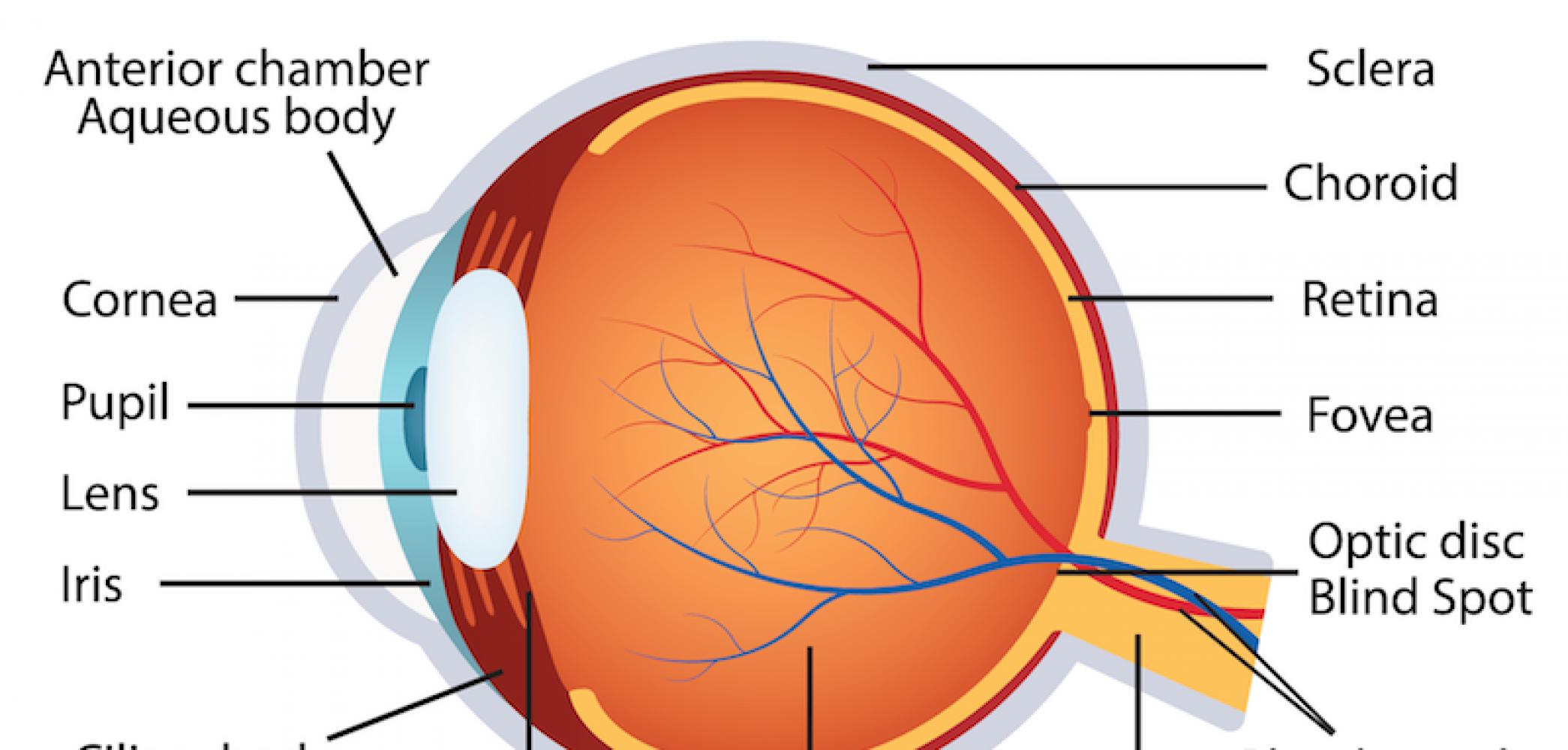
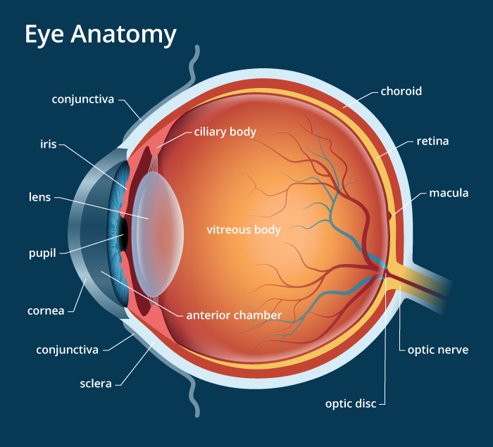
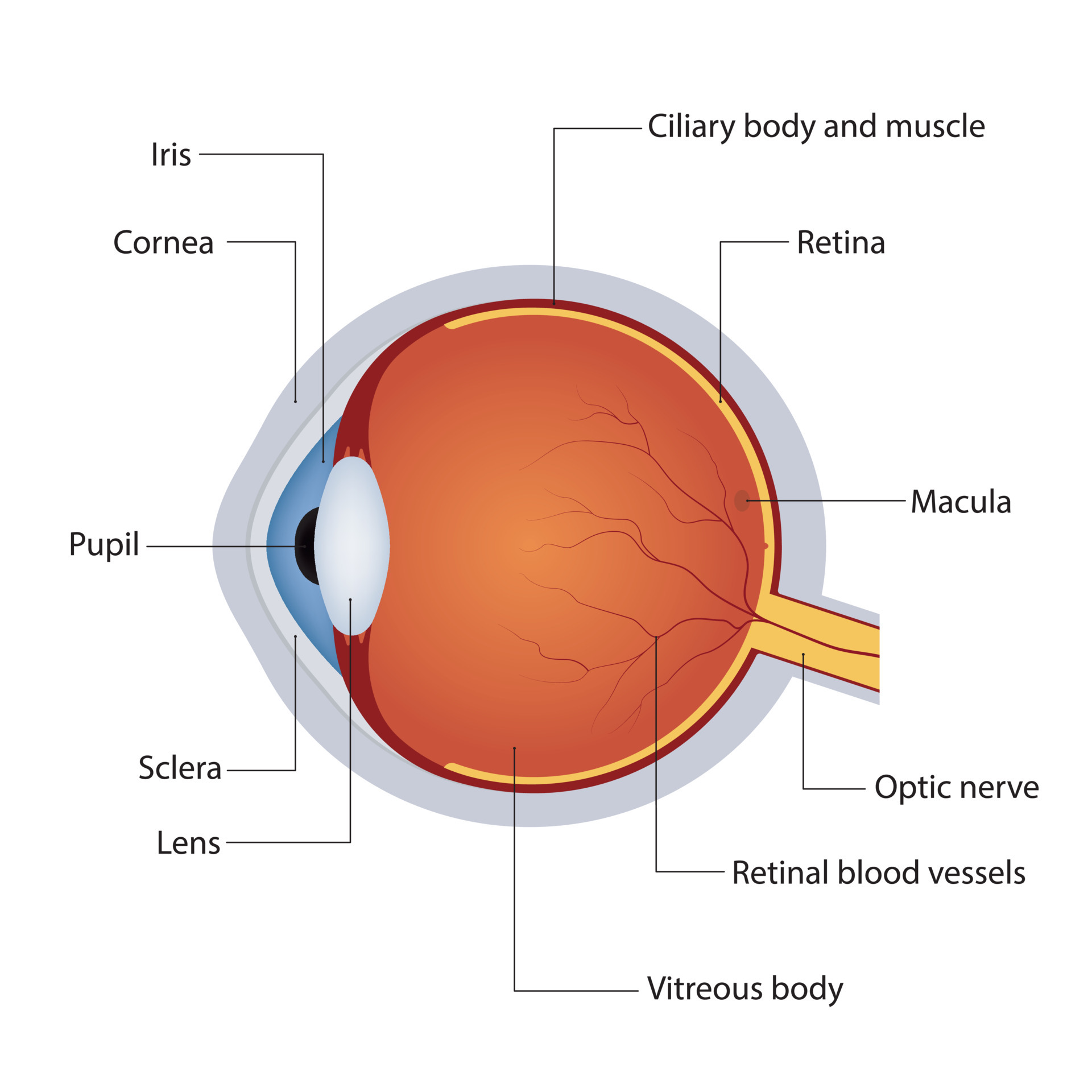
Human Eye Anatomy La Pine Eyecare Clinic Learn about the basic structure and function of the eye, from the outside to the inside. see diagrams and illustrations of the cornea, iris, lens, retina, optic nerve, and more. Learn about an ophthalmologist's role in eye care. get ophthalmologist reviewed tips and information about eye health and preserving your vision. learn about eye anatomy and learn how your eyes work with ophthalmologist approved facts.

Human Eye Anatomy Parts And Structure Online Biology Notes Learn how the eye works like a digital camera and see diagrams of its main structures, such as the cornea, iris, lens, retina and optic nerve. find out how vision problems affect your eyesight and how to protect your eyes. The following are parts of the human eyes and their functions: 1. conjunctiva. the conjunctiva is the membrane covering the sclera (white portion of your eye). the conjunctiva also covers the interior of your eyelids. the conjunctiva helps lubricate the eyes by generating mucus and tears. it also aids in immunological monitoring and prevents. Bony cavity within the skull that houses the eye and its associated structures (muscles of the eye, eyelid, periorbital fat, lacrimal apparatus) bones of the orbit. maxilla, zygomatic bone, frontal bone, ethmoid bone, lacrimal bone, sphenoid bone and palatine bone. structure of the eye. cornea, anterior chamber, lens, vitreous chamber and retina. Ciliary body. the part of the eye that produces aqueous humor. cornea. the clear, dome shaped surface that covers the front of the eye. iris. the colored part of the eye. the iris is partly responsible for regulating the amount of light permitted to enter the eye. lens (also called crystalline lens).

Structure Of Anatomy Human Eye Detailed Diagram Of Eyeball Side View Vector Illustration Bony cavity within the skull that houses the eye and its associated structures (muscles of the eye, eyelid, periorbital fat, lacrimal apparatus) bones of the orbit. maxilla, zygomatic bone, frontal bone, ethmoid bone, lacrimal bone, sphenoid bone and palatine bone. structure of the eye. cornea, anterior chamber, lens, vitreous chamber and retina. Ciliary body. the part of the eye that produces aqueous humor. cornea. the clear, dome shaped surface that covers the front of the eye. iris. the colored part of the eye. the iris is partly responsible for regulating the amount of light permitted to enter the eye. lens (also called crystalline lens). Learn about the anatomy and functions of the eye, as well as common eye conditions, tests, and treatments. see a diagram of the eye and its parts, such as iris, pupil, cornea, and retina. Human eye, specialized sense organ in humans that is capable of receiving visual images, which are relayed to the brain. the anatomy of the eye includes auxiliary structures, such as the bony eye socket and extraocular muscles, as well as the structures of the eye itself, such as the lens and the retina.

Ocular Anatomy Diagram Learn about the anatomy and functions of the eye, as well as common eye conditions, tests, and treatments. see a diagram of the eye and its parts, such as iris, pupil, cornea, and retina. Human eye, specialized sense organ in humans that is capable of receiving visual images, which are relayed to the brain. the anatomy of the eye includes auxiliary structures, such as the bony eye socket and extraocular muscles, as well as the structures of the eye itself, such as the lens and the retina.

Comments are closed.